
Journal of Health and Medical Sciences
ISSN 2622-7258


Published: 04 June 2026
Potential Expression of HER2 Protein According to the Macroscopic Type of Tumor in Gastric Cancer: Original Scientific Paper
Amir Spahić
Hospital Travnik, Bosnia and Herzegovina
Download Full-Text Pdf
10.31014/aior.1994.09.02.255
Pages: 35-48
Keywords: HER2, Macroscopic Type, Borrman, Gastric Carcinoma
Abstract
The aim of the study was to determine the relationship between HER2 protein expression and macroscopic tumor type according to Borrmann in gastric cancer. Method: This study used archival tissue samples of gastric cancer from 60 patients obtained after subtotal or total gastrectomy with regional lymphadenectomy of lymph nodes. The most representative tissue samples of gastric cancer tissue selected by the standard hematoxylin-eosin staining method were re-cut on a microtome and immunohistochemically treated with an antibody to HER2 according to the manufacturer's protocol, and then evaluated by light microscopy and scored using a modified scoring system for HER2 positivity of surgical resections. Descriptive statistics, non-parametric significance tests, namely Chi square test and One sample T-test, as well as correlation tests, namely Pearson's correlation test, were used in data processing. Results: Statistical analysis of HER2 protein expression according to the macroscopic appearance of gastric carcinoma according to Borrmann performed using the chi-square test does not show a statistically significant relationship between these two variables (χ2=66.267), analysis using the one sample T-test does not show a statistically significant difference (t=3.080), and analysis using the Pearson correlation test shows a positive correlation that is not statistically significant (r=0.069) between these two examined parameters. Conclusion: HER2 protein expression showed a negligible positive correlation without statistical significance according to the macroscopic type of carcinoma according to Borrmann using the univariate statistical method according to Pearson.
1. Introduction
According to the definition of the World Health Organization (WHO), gastric cancer is a malignant epithelial tumor of the gastric mucosa with glandular differentiation, and it is one of the most common tumors, and the second leading cause of death from cancer in the world, although its frequency has been declining, especially in developed countries, since the middle of the 20th century (Kelley and Duggan, 2003). The incidence of gastric cancer largely depends on the geographical area. It is especially common in the countries of the Far East, Asian countries of the former Soviet Union, Eastern European countries and South America. The world's highest incidence of this tumor is in Japan, where it is up to 80/100,000 inhabitants per year with about 100,000 newly diagnosed patients per year and is eight to ten times higher than in Western Europe, where it is 10 to 15 new cases per 100,000 inhabitants per year (Troskot and Gamulin, 2006). Tumor stage assessment is based on the basic principles of tumor growth and spread. In the initial phase of growth, the tumor spreads in the organ in which it originated, and with further growth it can directly spread to neighboring organs. During tumor growth, tumor cells enter blood and lymph vessels and spread (metastasize) to lymph nodes and distant organs. Therefore, the most common elements used in tumor stage assessment are:
1. location of the tumor inside the stomach (there is a special scheme that is used to assess the expansion of the tumor),
2. the depth of tumor invasion in the stomach wall,
3. the presence of tumors in the lymph nodes and the number of affected lymph nodes,
4. presence of distant metastases.
When analyzing gastric adenocarcinoma, in addition to determining the histological type according to Lauren and the macroscopic type according to Borrmann, it is necessary to determine the degree of differentiation, and Goseki grade (Gullick, 2001). Borrmann's macroscopic classification of advanced gastric carcinomas that invade muscle dates back to 1928 and distinguishes:
I: Polypoid, III: Ulcerated,
II: Fungal, IV: Infiltrative ("linitis plastica")
Types I, II/III, and IV tend to correspond to tubulo/papillary, intestinal, and adenocarcinomas of the "signet ring" cell type (linitis plastica), but there may be overlap between the categories. Polypoid/ulcerated tumors are considered to have a better prognosis than tumors of the infiltrative type. The tumor borders may be clear or unclear in relation to the surrounding tissue (Guan et al, 2009).
2. Molecular prognostic parameters
Overexpression of c-erbB-2 protein in gastric cancer (occurring in about 20% of cancer cases) has been shown to be an indicator of poor prognosis of gastric cancer (Jorgensen, 2010). However, such cancer cases may respond favorably to combination chemotherapy and trastuzumab (targeted therapy) (Hofmann et al, 2008).
Speaking of the p53 protein, in one series of studies from England (Gabbert et al, 2008) and another series of studies from Portugal (Allgayer et al, 1997), gastric cancers expressing the TP53 gene product were associated with a reduced survival rate (Dohchin et al, 2000), but in a series of studies from Germany such a relationship was not confirmed (Sgambato et al, 2000).
Also, increased levels of immunohistochemical detection of cathepsin D are associated with a worse survival rate in gastric cancer (Capuzzi et al, 2000). Similarly, high expression of cathepsin B and cathepsin L is associated with a greater propensity for tumor invasion and metastasis (Chung et al, 1996).
Immunohistochemical detection of the cyclin-dependent kinase p27 Kip1 has been demonstrated to be an indicator of reduced survival rates in patients with gastric cancer (Song et al, 2009). Preliminary studies indicate that loss of FHIT protein, or Bis (5-adenosyl)-triphosphatase, is an indicator of poor prognosis in gastric cancer (VanBeek et al, 2006). Expression of T-antigen as a precursor of the blood MN system is reported to correlate with the depth of invasion and the tendency of gastric cancer to metastasize to distant organs (Akiyama et al, 1986). EBV-positive gastric cancers have a better prognosis, probably due to the presence of activated cytotoxic T-cell infiltrate (Gullick, 2001; Coussens et al, 1985).
HER2 is a transmembrane glycoprotein essential for growth factor signaling. HER2 expression has also been found in colon, bladder, ovarian, endometrial, lung, cervical, head and neck, esophageal, and gastric cancers. Increased expression correlates with clinical outcome, conferring a poor prognosis, and is also a predictive factor for poor response to chemotherapy and endocrine therapy. The greatest value of determining HER2 status lies in predicting response to HerceptinR-Trastuzumab therapy (HERCEPTIN F.Hoffmann-La Roche Ltd, Basel, Switzerland and Genetech, Inc., South San Francisco, CA), in the sense that the efficacy of anti-HER2 therapy correlates with the degree of HER2 positivity, and Herceptin therapy has revolutionized the treatment of breast cancer (Hetzel et al, 1992; Hirashima et al, 2001; Douglass and Nava, 1985).
The TNM classification is currently the most important prognostic factor for gastric cancer. However, the prognosis in different patients with the same stage of the disease is not the same. Therefore, it is necessary, in addition to the TNM classification and the classic pathological characteristics of the tumor, to identify biological prognostic factors, which are often derived from genetic processes and are considered to represent a key step in the prognosis of gastric cancer.
There are opinions that HER2 expression plays a key role in the development of gastric cancer, its progression and metastasis. However, little is known about its expression, because a limited number of papers have been published on this topic in the world (Gravalos and Jimeno, 2008). Given that the majority of stomach cancers, nowadays, unfortunately, are diagnosed at an advanced stage of the disease, it is still unclear whether the expression of the HER2 protein in this cancer is related to any of the proven prognostic factors, and whether it can have a potential role in the prognosis of the disease and possibly provide guidelines for therapeutic treatment.
The aim of the work was to determine the correlation of HER2 protein expression with macroscopic tumor type according to Borrmann in gastric cancer.
2. Material and methods
The research conducted and presented in this paper represents a fundamental or basic type of research with an observational method of obtaining data used in the study.
2.1. Material
This study used archival tissue samples of gastric carcinoma from 60 patients obtained after subtotal or total gastrectomy with regional lymphadenectomy of the lymph nodes of the perigastric fatty tissue along the lesser and greater curvature of the stomach, then the lymph nodes of the celiac tripus region and the hepatoduodenal ligament region. Pathohistological diagnostics of all gastric carcinoma samples included in this study was performed at the Department of Pathology and Cytodiagnostics of the Travnik Hospital, in the period from the beginning of 2010 to the end of 2012. The condition for including samples in this study is that the patients had an operable tumor with a known status of regional lymph nodes and without evident distant dissemination at the time of diagnosis. The same number, i.e. 60 samples of normal stomach tissue, were used as a control group. In the prospective part, a pathohistological profile of HER2 amplifying gastric carcinomas was constructed based on a retrospective study, which was applied in the selection of pathohistologically verified gastric carcinomas in which immunohistochemical detection of HER2 protein was performed. Data on the clinicopathological parameter of the macroscopic tumor type according to Borrmann were obtained by macroscopic examination of the material after subtotal or total gastrectomy.
2.2. Methods
Pathohistological processing of biopsy material obtained by subtotal or total gastrectomy with regional lymphadenectomy involved taking tissue samples according to dissection rules, and their standard histochemical processing and, in the first act, staining with the standard hematoxylin-eosin method as well as with the special histochemical method of staining PAS (Periodic Acid Schiff) for mucin. In the next procedure, the most representative tissue samples of gastric carcinoma tissue embedded in paraffin blocks were re-cut on a microtome and immunohistochemically stained, i.e. treated with an antibody to HER2 according to the manufacturer's protocol, and then evaluated by light microscopy and scored using a modified HER2 positivity scoring system for surgical resections.
2.3. Guide to HercepTest scoring in gastric carcinoma
Tests to test the consistency, i.e. validity of the results between immunohistochemical staining and in situ hybridization of the pre-ToGA study showed that the HER2 IHC test is suitable for staining gastric carcinoma cells, but differences appeared compared to breast carcinoma, and changes were required in the scoring system for gastric carcinoma. The pre-ToGA and ToGA (Trastuzumab for Gastric Cancer) studies showed that a modified HER2 positivity scoring system should be used for gastric carcinoma samples.
2.4. Ethical aspects of the study
The use of archival gastric cancer tissue samples in this study was approved by the Ethics Committee of the Travnik Hospital, the institution where the study was conducted, for the purpose of using the data for scientific purposes.
2.5. Statistical data processing
After microscopic processing of the material, the obtained data were entered into MS Excel 2007. The data stored in this way were transported to the SPSS 16.0 software package for statistical processing. Descriptive statistics, non-parametric significance tests, namely the Chi square test and One sample T-test, as well as correlation tests, namely the Pearson correlation test, were used in data processing.
3. Results
Analyzing the macroscopic appearance of the cancer tissue of the samples of the experimental group and using Borrmann's macroscopic classification, the results obtained showed that 4 samples, or 6.7% of the total number of samples of the experimental group, had a macroscopic appearance of a polypoid tumor mass, while only one sample had a fungal macroscopic appearance, which is 1.7% of the total number of samples of the experimental group. Furthermore, 41 samples, or 68.3% of the total number of samples, had the appearance of an ulcerative lesion, while a total of 14 samples, or 23.3% of the total number of samples of the experimental group, showed an infiltrative type of macroscopic appearance of the tumor. Data on the prevalence of individual macroscopic forms of gastric cancer according to Borrmann are numerically and in percentages shown in Chart 1.

After processing and analyzing the characteristics of the macroscopic tumor type according to Borrmann of the cancer samples of the studied group of patients, immunohistochemical staining and analysis of all samples of the experimental and control groups of samples are performed.
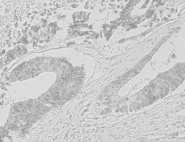
The immunohistochemical analysis of the application of Herceptest on the experimental group of gastric cancer biopsy samples obtained results showing that the largest number of gastric cancers showed HER2 negative immunoreactivity, that is, that the largest number of gastric cancers did not show immunoreactivity in the expression of the HER2 protein, and out of a total of 60 samples that made up the experimental group, 36 of them, or 60%, showed HER2 negative immunoreactivity (Figure 1). Furthermore, weak or barely visible membrane activity, some of which are only in parts of the cell membranes of the cancer cells, is shown by a total of 9 samples, or 15% of the total number of samples of the experimental group (Figure 2).
Ambiguous membrane activity was shown by 8 samples of the experimental group, i.e. 13.33% of the total number of samples of the experimental group (Figure 3), and these samples in further work should be retested by the method of in situ hybridization in the light microscopic field (CISH).
And finally, what is most important and why the research was done, hyperreactivity, i.e. baseolateral or lateral membrane reactivity in HER2 protein expression, was shown by 7 samples, i.e. 11.67% of the total number of samples of the experimental group (Figure 4).
One sample showed apical membranous activity, with no evident baseolateral or lateral membranous activity in HER2 protein expression, and that sample was considered a HER2-negative immunoreactivity sample (Figure 5).
All samples from the control group of samples, which were represented by samples from histologically regular stomachs, showed HER2 negative immunoreactivity (Figure 6).
Examining the relationship between Borrmann's classification of macroscopic appearances of gastric carcinoma and membranous immunoreactivity in HER2 protein expression, the results showed that as many as 6 samples with hyperreactivity in HER2 protein expression, or 10% of the total number of samples in the experimental group, had a macroscopic appearance of an ulcerative lesion, while 1 sample with hyperreactivity, or 1.7% of the total number of samples, had the appearance of a diffuse infiltrative lesion according to Borrmann. Ambiguous membranous activity was shown by 5 samples, or 8.3% of the total number of samples with the appearance of an ulcerative lesion, and 3 samples, or 5% of the total number of samples with the appearance of a diffuse infiltrative lesion. Gastric carcinoma lesions with the appearance of polypous and fungal lesions did not show either hyperreactivity or ambiguous membrane activity in HER2 protein expression, which is numerically and in percentages shown in graph 2.
The control group samples did not show immunoreactivity in HER2 protein expression, and they do not have a defined macroscopic appearance according to Borrmann.

Statistical analysis of HER2 protein expression according to the macroscopic appearance of gastric cancer according to Borrmann performed using the chi-square test does not show a statistically significant relationship between these two variables (χ2=66.267; a statistically significant relationship is if p<0.05), analysis using the one sample T-test does not show a statistically significant difference (t=3.080; the difference is not statistically significant if p>0.05), and analysis using the Pearson correlation test shows a positive correlation that is not statistically significant (r=0.069) between these two examined parameters.
4. Discussion and conclusion
The aim of the study was to qualitatively and quantitatively determine the possible expression of HER2 protein in gastric cancer, and to examine the correlation of HER2 protein expression with the examined clinicopathological parameter of gastric cancer, i.e. the macroscopic tumor type according to Borrmann.
Before discussing the main objectives of this study, we need to discuss the basic characteristics, i.e. the clinicopathological parameter of the experimental research group.
Analyzing the macroscopic appearance of the carcinoma samples of the experimental research group using Borrmann's macroscopic classification from 1928, we obtained results that showed that 4 samples, i.e. 6.7% of the total number of samples of the experimental group, had the macroscopic appearance of a polypoid tumor mass, while only one sample had a fungal macroscopic appearance, which is 1.7% of the total number of samples of the experimental group. Furthermore, 41 samples, which is 68.3% of the total number of samples, had the appearance of an ulcerative lesion, while a total of 14 samples, which is 23.3% of the total number of samples of the experimental group, showed the infiltrative type of macroscopic tumor appearance.
Examining the relationship between Borrmann's division of the macroscopic appearance of gastric cancer and membrane immunoreactivity in the expression of the HER2 protein, the result was that as many as 6 samples with hyperreactivity in the expression of the HER2 protein, i.e. 10% of the total number of samples of the experimental group, had the macroscopic appearance of an ulcerative lesion, while 1 sample with hyperreactivity, which was 1.7% of the total number of samples, had the appearance of a diffuse infiltrative lesion according to Borrmann. Ambiguous membrane activity was shown by 5 samples or 8.3% of the total number of samples with the appearance of an ulcerative lesion and 3 samples or 5% of the total number of samples with the appearance of a diffuse infiltrative lesion. Gastric carcinoma lesions with polypous and fungal appearance showed neither hyperreactivity nor ambiguous membrane activity in HER2 protein expression. Weak membrane activity in HER2 protein expression was shown by 1 gastric carcinoma sample with polypous appearance, then fungal and diffuse infiltrative lesions, which is 1.7% of the total number of samples, and 6 carcinoma samples with macroscopic appearance of ulcerative lesions, which is exactly 10% of the total number of samples in the experimental group. Negative HER2 immunoreactivity was shown by 3 polypous-appearing carcinoma samples, which is exactly 5% of the total number of samples, then 24 ulcerous-appearing carcinoma samples, which is exactly 40% of the total number of samples, and 9 diffuse infiltrative carcinoma samples, which is exactly 15% of the total number of samples in the experimental group. Control group samples did not show immunoreactivity in HER2 protein expression, and do not have a defined macroscopic appearance according to Borrmann. Statistical analysis of HER2 protein expression according to the macroscopic appearance of gastric carcinoma according to Borrmann performed using the chi-square test does not show a statistically significant relationship between these two variables (χ2=66.267; statistically significant relationship is if p<0.05), analysis using the one sample T-test does not show a statistically significant difference (t=3.080; the difference is not statistically significant if p>0.05), and analysis using the Pearson correlation test shows a positive correlation that is not statistically significant (r=0.069) between these two examined parameters.
The frequencies of HER2 protein expression in published studies vary enormously from 8% to 91%. One of the first studies on this topic was performed in 2000 when Allgayer et al. examining the frequency of HER2 protein expression in gastric carcinoma obtained an extremely high expression of 91%. However, both in this and other studies originally conducted on this topic, tumor heterogeneity was neglected, which is extremely pronounced in gastric cancer compared to breast cancer (4.80%:1.40%), and the malignant cell scoring system used was for breast cancer. These data can be taken as the reason for such a high percentage of HER2-positive gastric cancers (Heike et al, 2010).
Newer studies conducted after the pre-ToGa and large multicenter ToGa studies took into account the heterogeneity of gastric cancer, as well as a modified tumor cell scoring system adapted for gastric cancer. In our study, these two parameters were also taken into account and we obtained 11.67% of HER2-positive cancers, which is similar and different from the findings of previous studies: the large multicenter ToGa (Trastuzumab for GAstric Cancer) study in which there were 22.10%, the results of the Chinese study conducted by Guan Zhen Yu et al. who had overexpression of HER2 protein in 28% of the primary gastric adenocarcinomas examined, as well as the results of the largest, most recent, study conducted to date on 924 patients in which 7% of the cancers were HER2 positive (Guan et al, 2009).
In our study, a negligible positive correlation without statistical significance of HER2 protein expression was demonstrated by univariate statistical method in the macroscopic appearance of the cancer (Siewert et al, 1998).
Funding: Not applicable.
Conflict of Interest: The authors declare no conflict of interest.
Informed Consent Statement/Ethics Approval: Not applicable.
Declaration of Generative AI and AI-assisted Technologies: This study has not used any generative AI tools or technologies in the preparation of this manuscript.
References
Akiyama T, Sudo C, Ogawara H. The product of the human c-erbB–2 gene: a 185-kilo-Dalton glycoprotein with tyrosine kinase activity 1986; 232: 1644–6.
Allgayer H, Babic R, Grützner KU, Beyer BC, Tarabichi A, Schildberg FW, et al. An immunohistochemical assessment of cathepsin D in gastric carcinoma: its impact on clinical prognosis. Cancer 1997; 80: 179-87.
Capuzzi D, Santoro E, Hauck WW, Kovatich AJ, Rosato FE, Baffa R, et al. A fhit expression in gastric adenocarcinoma: correlation with disease stage and survival. Cancer 2000; 88: 24-34.
Chung YS, Yamashita Y, Kato Y, Nakata B, Sawada T, Sowa M. Prognostic significance of T antigen expression in patients with gastric carcinoma. Cancer 1996; 77: 1768-73.
Coussens L, Yang-Feng TL, Liao YC. Tyrosine kinase receptor with extensive homology to EGF receptor shares chromosomal location with neu oncogene 1985; 230: 1132–9.
Dohchin A, Suzuki J, Seki H, Masutani M, Shiroto H, Kawakami Y. Immunostained cathepsins B and L correlate with depth of invasion and different metastatic pathways in early stage gastric carcinoma. Cancer 2000; 89: 482-7.
Douglass Jr HO, Nava HR. Gastric adenocarcinoma. Management of the primary disease. Semin Oncol 1985; 12: 32-45.
Gabbert HE, Müller W, Schneiders A, Meier S, Hommel G. The relationship of p53 expression to the prognosis of 418 patients with gastric carcinoma. Cancer 1995; 76: 720-6.
Gravalos C, Jimeno A. HER2 in gastric cancer: a new prognostic factor and a novel therapeutic target. Annals of Oncology 2008;19(9): 1523–9.
Guan ZY, Ying C, Jie JW. Overexpression of Grb2/HER2 signaling in Chinese gastric cancer: their relationship with clinicopathological parameters and prognostic signifcance. J Cancer Res Clin Oncol 2009; 135: 1331–9.
Guan ZY, Ying C, Jie JW. Overexpression of Grb2/HER2 signaling in Chinese gastric cancer: their relationship with clinicopathological parameters and prognostic signifcance. J Cancer Res Clin Oncol 2009; 135: 1331–9.
Gullick WJ. Update on HER-2 as a target for cancer therapy: alternative strategies for targeting the epidermal growth factor system in cancer. Breast Cancer Res 2001; 3: 390-4.
Gullick WJ. Update on HER-2 as a target for cancer therapy: alternative strategies for targeting the epidermal growth factor system in cancer. Breast Cancer Res 2001; 3: 390-4.
Heike G, Shivan S, Sally G, Helmut E. Gabbert and Wolfram Müller HER2 expression in gastric cancer: Rare, heterogeneous and of no prognostic value – conclusions from 924 cases of two independent series. Cellular Oncology 2010; 32: 57–65.
Hetzel DJ, Wilson TO, Keeney GL. HER-2/neu expression: a major prognostic factor in endometrial cancer. Gynecol Oncol 1992; 47: 179–85.
Hirashima N, Takahashi W, Yoshii S. Protein overexpression and gene amplification of c-erb B-2 in pulmonary carcinomas: a comparative immunohistochemical and fluorescence in situ hybridization study. Mod Pathol 2001; 14: 556–662.
Hofmann M, Stoss O, Shi D, Buttner R, Kim W, Ochiai A, et al. Assessment of a HER2 scoring system for gastric cancer: results from a validation study. Histopathology 2008; 52: 797-805.
Jorgensen JT. Targeted HER2 treatment in advanced gastric cancer. Oncology 2010; 78: 26-33.
Kelley JR, Duggan JM. Gastric cancer epidemiology and risk factors. J Clin Epidemiol 2003; 56:1-9.
Sgambato A, Migaldi M, Leocata P, Ventura L, Criscuolo M, DiGiacomo C, et al. Loss of p27 KIP1 expression is a strong independent prognostic factor of reduced survival in N0 gastric carcinomas. Cancer 2000; 89: 2247-57.
Siewert JR, Bottcher K, Stein HJ. Relevant prognostic factors in gastric cancer: ten-year results of the German Gastric Cancer Study. Ann Surg. 1998;228: 449–61.
Song HJ, Noh JH, Joo M, Jang KT, Park CK, Kim KM. The type of host inflammatory immune response predicts survival in EBV-associated gastric carcinomas: a multivariate analyses of 109 cases. Lab Invest 2009; 89(Suppl 1): 148A.
Troskot B, Gamulin M. Gastric Adenocarcinoma. Medicus 2006;15, 73 – 87.
VanBeek J, Snel SN, Berkhof J, Kranenbarg EK, Middeldorp JM, Meijer CJ, et al. Morphological vidence of an activated cytotoxic T-cell infiltrate in EBV-positive gastric carcinoma preventing lymph node metastases. Am J Surg Pathol 2006; 30: 59-65.